

About the Gallery....
Here and on the following Gallery pages, you will see more examples of blind deconvolutions performed using the SeDDaRA process. A great advantage of this technique is that it can be applied to any data type, such as consumer photographs, medical images, one dimensional signals, microscopy, and astronomy images. For most, the improvement is obvious. In some cases, the improvement more subtle. SeDDaRA removes the blur that is present in the image (or signal) by extracting the point spread function (or impulse function). Static images are below.
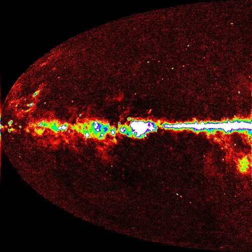
Quarktet's blind deconvolution algorithm, SeDDaRA, was named as one of Advanced Imaging Magazine's Solution of the Year way back in 2004. The winning solution is the removal of blur from an image of the background radiation of the universe. A portion of the image and its restoration appear below.
  |
|
Fig1: Image of the background radiation of the universe as taken by the NASA Wilkinson Microwave Anistropy Probe (WMAP) satellite. The image has been false-colored to highlight the contrast. This image, together with four images in four other spectral bands, allowed NASA/WMAP scientists to more accurately define the age of the universe and the rate of expansion. Fig 2: Using a fractal image as a model, a blur function, resembling a clover-leaf, was extracted from the image. A pseudo-inverse filter removed the blur to sharpen the image. The image is cleaner and reveals greater resolution. We hope the use of this processing technique will enable scientists to achieve even greater accuracy in their calculations. Image Courtesy of the NASA/WMAP Science Team. |
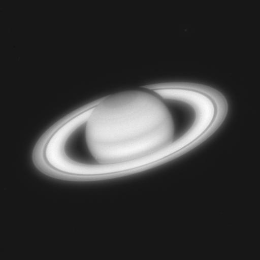

Fig1: Image of Saturn, taken by the Hubble Space Telescope before the optical correction was applied in December 1993. This image has become a standard image deconvolution example.
Fig 2: Restored Image. Not only is the planet much cleaner, but if you look close enough you will see stars behind the planet that were not apparent before.


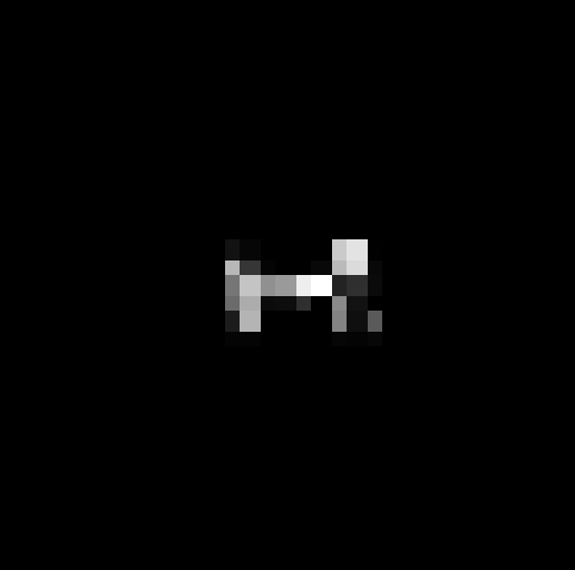
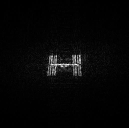
Fig1: A microscope image of fission yeast that already undergone some processing. This image was displayed on several websites as an example of good deconvolution.
Fig 2: Restoration of the microscope image. Even though the image has already been processed by more conventional means, the image is significantly enhanced using SeDDaRA. This occurs because it is very difficult to measure or calculate the exact blur function of a system. Many times, a little help is needed to see clearly.

